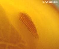
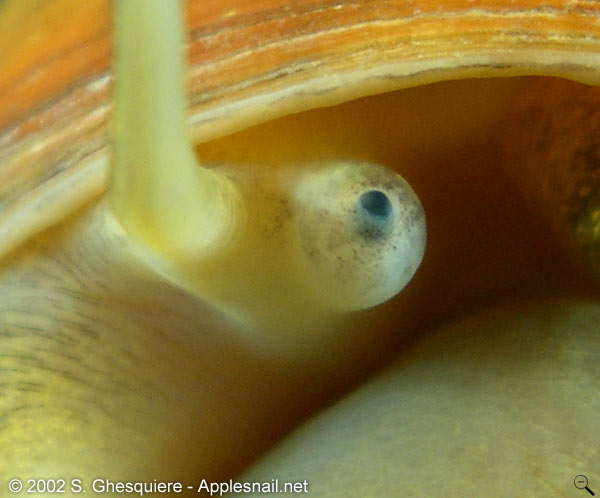
 Eye at the
base of a tentacle (Pomacea canaliculata).
Eye at the
base of a tentacle (Pomacea canaliculata).
(click on picture for large version)
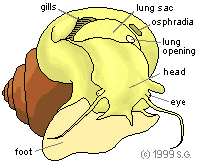
| Sensory system |
|
Overview 3D anatomy Digestion Senses Nerves Respiration Circulation Reproduction Other Shell |
|
|
Apple snails are highly depending on their smell sense. With their smell
sense they are able to locate food and recognise other snails of their own species, which
is important to find a good mating partner. It's therefore not surprising that apple snail
have a well-developed smell sense.
The vision of the apple snail on the other hand is rather weak (poor ability to form
images and bad image quality) and functions merely as a light direction detector.
The tactile sense is well developed as can be seen as the snail walks
over a small object or encounters an obstacle.
The hearing capabilities of the apple snail are worthless, even more: they are completely
deaf.
Body: The whole body surface of the apple snail contains chemo- and
mechanoreceptors.
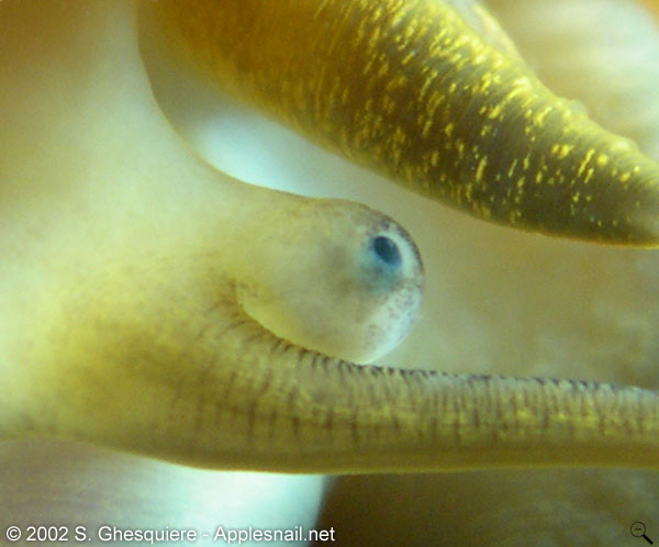
Eyes: The eyes of the apple snail are located at the
base of the tentacles, on top of the eyestalks.
The structure of the eye does not provide detailed vision, they rather function
as directional light sensors that give the snail an orientation towards light
sources. Colour vision is absent as the retina does not contain colour specific
photosensors: an apple snail is colour-blind.
| Movies (MPEG1): - The eyes of an apple snail (Asolene megastoma) (132kb) |
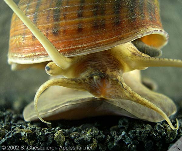
 The eye on the
eye stalk of a Pomacea canaliculata
snail. The eye on the
eye stalk of a Pomacea canaliculata
snail. (click on image to enlarge) |
 Another photograph
of an eye (Pomacea canaliculata). Another photograph
of an eye (Pomacea canaliculata).
(click on image to enlarge) |
It appears that the eyes of apple snails are rather optimized for optical
sensitivity (night vision!) rather than for high detail vision. In fact, two
mechanism are used in the apple snail eye to increase sensitivity to light:
the eyes are relatively large with a large lens (large eyes= more light captured)
and the light sensitive cells (rhabdoms) are grouped and connected with the
same neuron (pooling of signals or neural summation = higher sensitivety, but
lower resolution). It has even been suggested that the optical sensitivity of
the apple snail approaches that of the nocturnal spider Dinopis subrufus
(Blest and Land, 1977).
The spatial resolution of the eye, however, is estimated to be around a poor
44 pixels (SEYER, J.O. et al. 1998). This
low resolution is due to several factors: the pooled light sensitive cells,
the bad quality of the lens (blurring) and unisolated light sensitive cells
(high cross-talk between cells). To give an idea, human eyes are capable to
resolve 10.000.000 pixels. The pictures below illustrate this even better.
As apple snail do not have colour sensitive cells in their retina, they can
only see shades of grey (colour-blind).
One can conclude that the eyes of apple snails are suitable to find light and
dark area's within their invironment, even at night, but that their overal vision
in the sense of image quality is rather poor.
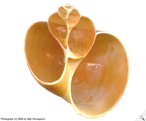 A photograph
of a Pomacea diffusa
shell, cut in half. A photograph
of a Pomacea diffusa
shell, cut in half. |
 The same photograph seen through the eyes of an apple snail. Simulated with blur from lens, pooled cells and cross-talk. Image as one would probably receive from the neural cells. |
The anatomy of the apple snail eye is simple and consists of several structures
embedded in the connective tissue of the eyestalk: the cornea, the lens, the
retina and the optic nerve.
The acellular lens, with a spherical shape, is embedded in the eyestalk. The lens itself
consists of a dense ovoid inner core with several peripheral layers at the outside.
The thin cornea on top of the eye forms a circular pupil. This transparant tissue
at which light enters the eye consists of 4 layers. The peripheral double layer
of the cornea has a cuboidal epithelium layer at the outside and a connective
tissue layer with a collagenous structure at the inside. The epithelium at the
outside is continuous with the epidermis covering the whole eyestalk and the
rest of the body.
The inside of the cornea is part of the lens and is a double layer as well, with
microvilli at the inside and an epithelial layer near the periphery.
The retina at the base of the lens is divided in four separate layers. The first layer,
surrounding the lens, contains light-sensitive microvillar rhabdoms. The second layer is
heavily pigmented and composed of the pigmented parts of the photoreceptor cells and
isolated pigment cells. Around this layer is a pigmented layer in which the nuclei of
photoreceptor cells are embedded together with supportive cells. The fourth, outermost
layer consist of mainly connective tissue containing neural cells and their axons, which
connect with the optic nerve.
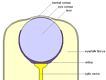 Overview illustration of the eye. |
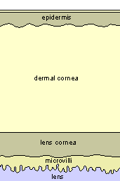 Close up of the cornea. |
Tentacles: The tentacles are very
important sensory organs. Apple snails highly rely on the smell capacity and the
sensitivity of their tentacles to navigate in their environment.
The cephalic tentacles (attached at the top of the head) can be very long, sometimes even
longer than the body of the snail.
The labial tentacles or labial palps (attached at each side of the mouth), a typical
feature of apple snails, are shorter.
Osphradia: The osphradius is a chemosensory
structure that is located in the mantle cavity, in front of the lung.
The osphradius gives the snail the capability to smell chemical substances in the water.
|
|
The statocysts: The statocysts (2) are
vesicles containing a statolith (little stone like structures composed of calcium
carbonate). They function as balancing organs, used by the snail to detect its position
with regard to the ground.
They are located inside the snail's body close to the pedal
ganglia.
|
|
|
|
Except where otherwise noted, this page is licensed under a Creative Commons Attribution-NonCommercial-ShareAlike 2.5 License . http://www.applesnail.net |
|